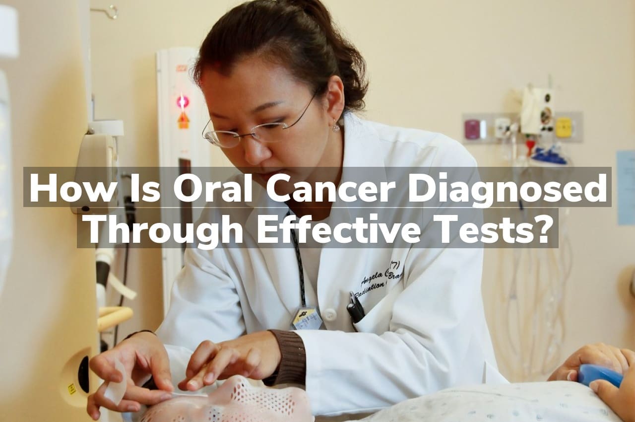
How is oral cancer diagnosed through effective tests? At Joel W. Yates Jr. D.D.S., we utilize a combination of thorough visual examinations and advanced diagnostic tools to identify signs of oral cancer early, ensuring prompt and precise intervention for the best possible outcomes.
Early Detection: Key Screening Methods
Early detection of oral cancer significantly increases the chances of successful treatment and survival. Dentists and doctors often perform routine oral examinations to look for signs of cancer, even before symptoms appear. This includes a visual inspection of the mouth, face, and neck for any abnormalities, as well as a physical palpation of these areas to feel for lumps or irregular tissue changes. For those at higher risk, such as tobacco and alcohol users, these screenings are particularly crucial. Additionally, newer technologies like VELscope, a special light that can help visualize changes in the oral mucosa, are enhancing early detection capabilities. For a more in-depth understanding of these procedures, consider Understanding the process of testing for oral cancer, which provides valuable insights into the various diagnostic tools used by healthcare professionals.
Aside from visual and tactile examinations, other screening methods include the use of toluidine blue dye, which can help identify suspicious areas, and brush biopsies that collect cells from the oral tissues for further analysis. When any suspicious lesions are found, a definitive diagnosis is usually made through a scalpel biopsy, where a small piece of tissue is removed and examined under a microscope. It’s important to note that not all irregularities turn out to be cancerous, but early screening and testing are essential steps in ensuring that if oral cancer is present, it is diagnosed at an early stage when treatment is most likely to be effective.
Biopsy: The Definitive Diagnostic Tool
A biopsy is often the most conclusive method for diagnosing oral cancer. During this procedure, a small sample of tissue is removed from the suspicious area and examined under a microscope by a pathologist. This test can determine whether the cells are cancerous and, if so, the type and grade of the cancer. There are different types of biopsies, including incisional, where only a portion of the tissue is removed, and excisional, where the entire lesion is excised. The choice of biopsy depends on the size and location of the abnormal area.
A biopsy not only confirms the presence of cancer but can also provide vital information about the extent of the disease and guide treatment decisions. For individuals seeking comprehensive oral cancer evaluations, Jefferson Oral Cancer Screening Services offer a range of diagnostic options, including biopsies performed by experienced professionals. Early detection and accurate diagnosis are critical in managing oral cancer effectively, and these services play a pivotal role in patient care.
Imaging Techniques: Beyond the Naked Eye
Advancements in imaging techniques have revolutionized the early detection and diagnosis of oral cancer, offering a glimpse beyond what the naked eye can see. These non-invasive methods, such as X-rays, CT scans, MRI, and PET scans, allow healthcare professionals to detect abnormalities and assess the extent of cancer with remarkable precision. X-rays can reveal hidden tumors in the jaw and neck, while CT scans provide detailed cross-sectional images that help in evaluating the tumor’s size and location.
MRI scans offer a more detailed view of soft tissues, including lymph nodes and muscles, aiding in determining cancer’s stage. PET scans are particularly effective in identifying cancer spread, as they can detect areas of increased metabolic activity typical of cancer cells. By integrating these imaging techniques into the diagnostic process, doctors can formulate a more accurate and tailored treatment plan, improving patient outcomes and survival rates.
Brush Biopsy: A Non-Invasive Approach
When it comes to the early detection of oral cancer, a Brush Biopsy is a non-invasive diagnostic tool that stands out for its simplicity and comfort. This procedure involves the gentle brushing of the oral lesion to collect cells for analysis, without the need for scalpel or anesthesia, making it an ideal preliminary test. The collected cells are then examined under a microscope to identify any abnormal changes or signs of malignancy.
The ease and minimal discomfort of a brush biopsy encourage patients to undergo regular screenings, thereby increasing the chances of early detection and successful treatment of oral cancer. With its straightforward application, a brush biopsy serves as a crucial step in the fight against this serious disease, offering a quick and effective method for healthcare professionals to assess oral health concerns.
HPV Testing: Oral Cancer’s Viral Link
Human Papillomavirus (HPV) has emerged as a significant factor in the development of oral cancer, particularly oropharyngeal cancers affecting the back of the throat, base of the tongue, and tonsils. HPV testing plays a crucial role in the early diagnosis of oral cancer, as it can identify the presence of high-risk HPV strains known to contribute to cancerous changes in oral tissues. This test is typically performed on a sample of cells taken from a suspected lesion or during a routine dental examination.
A positive HPV test indicates that a patient may be at an increased risk for developing oral cancer, prompting further investigation and potentially more aggressive monitoring or treatment. As awareness of HPV’s link to oral cancer grows, HPV testing has become an integral part of the diagnostic process, aiding healthcare professionals in identifying at-risk individuals and implementing timely interventions.
Conclusion
For a thorough evaluation and to discuss oral cancer screening, call Joel W Yates Jr. D.D.S at 336-846-2323, or read our reviews on Google Maps.


